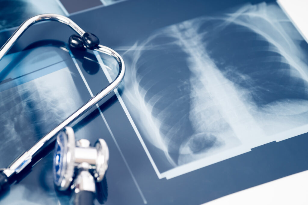
At Behan Orthopedics, we understand the importance of accurate diagnostics in your treatment journey. Our state-of-the-art X-Ray Services in Bay City, Michigan, are designed to provide detailed images that help our expert orthopedic doctors diagnose and treat your musculoskeletal disorders effectively.
Our advanced X-ray technology ensures precise imaging, leading to accurate diagnoses and effective treatment plans.
Our orthopedic team in Bay City, Michigan, is highly experienced and dedicated to providing the highest standard of care.
We treat every patient with empathy and respect, ensuring you feel comfortable and supported throughout your treatment journey.
X-rays are a form of electromagnetic radiation that can pass through the body to create images of its internal structures.
By understanding how X-rays work and their benefits, you can approach your diagnostic journey with confidence. At Behan Orthopedics, we’re committed to using the latest X-ray technology to provide you with the most accurate diagnoses and effective treatment plans.
X-rays are produced by an X-ray machine that sends a controlled amount of radiation through the body. Different tissues absorb X-rays at different rates. Dense materials, like bone, absorb more X-rays and appear white on the X-ray film. Softer tissues, like muscles and organs, absorb fewer X-rays and appear in shades of gray.
Standard X-Rays: Often used to diagnose broken bones, infections, and abnormalities in the chest and abdomen.
Computed Tomography (CT) Scans: Provide more detailed images by taking multiple X-rays from different angles and creating cross-sectional views.
Fluoroscopy: Uses a continuous X-ray beam to create real-time images, often used during procedures like joint injections and catheter insertions.
Digital Radiography: Offers immediate image viewing and can be easily stored and shared electronically.
Non-invasive: X-ray imaging is painless and doesn’t require any incision.
Quick Results: X-rays provide fast results, which are crucial in emergency situations.
Versatile: Can be used to diagnose a wide range of conditions, from fractures and infections to tumors and arthritis.
Guidance for Procedures: Helps guide orthopedic surgeons during complex procedures to ensure precision.
While X-rays involve exposure to radiation, the amount used is generally very low and considered safe for most patients. Nevertheless, special precautions are taken, especially for pregnant women and children, to minimize any potential risks.
Minimizing Exposure: Lead aprons and other protective measures are used to shield parts of the body not being imaged.
Informed Consent: Patients are informed about the benefits and risks to make an educated decision about their care.
Remove Jewelry: Metal objects can interfere with the image quality.
Follow Instructions: Depending on the type of X-ray, you may need to fast or take other preparatory steps.
Stay Still: Remaining still during the imaging process ensures clear and accurate results.

Your health and well-being are our top priorities. Whether you’re dealing with a sports injury, arthritis, or any musculoskeletal disorder, Behan Orthopedics is here to help.
Insurances may be subject to certain plans or authorizations.


